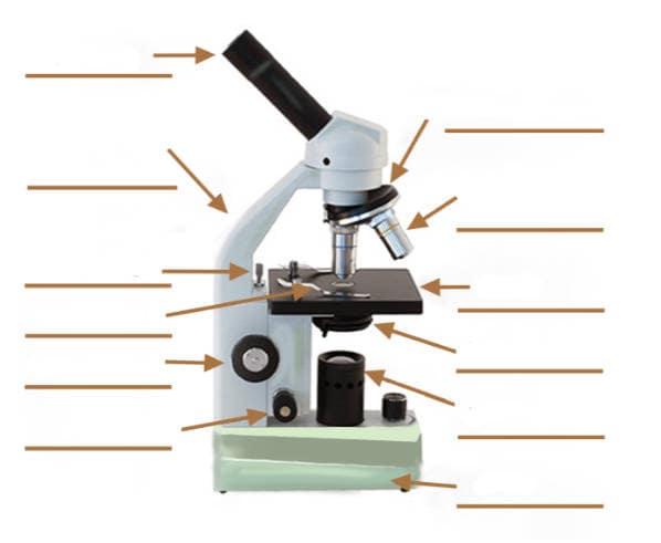
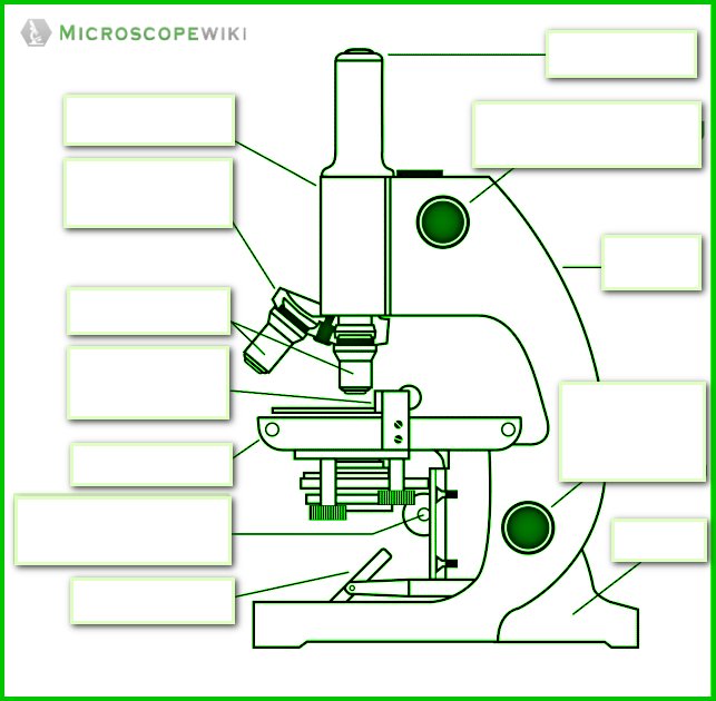
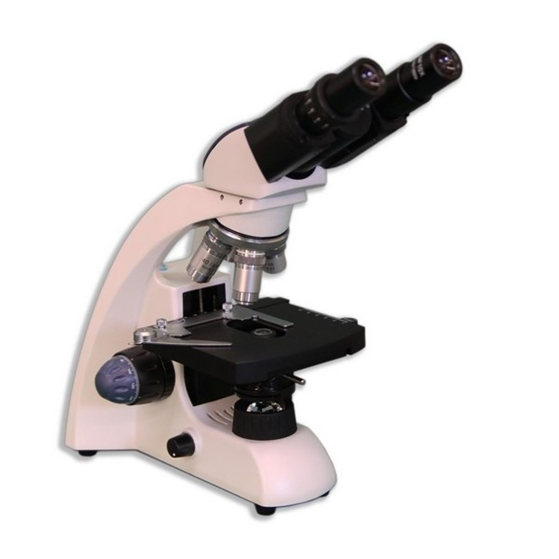
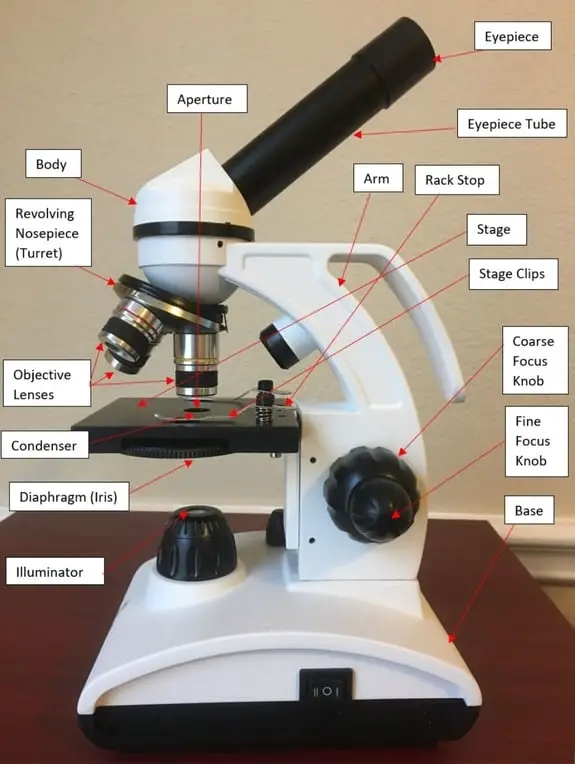
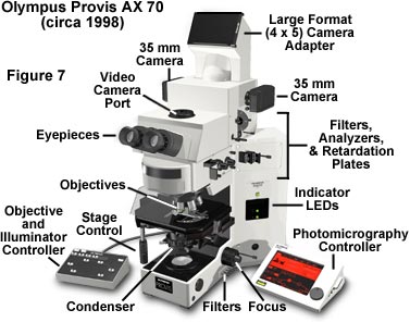
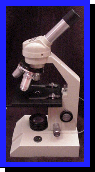
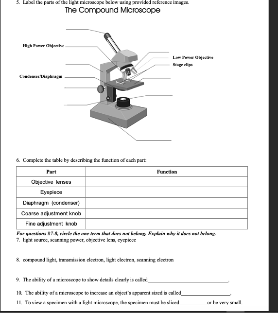
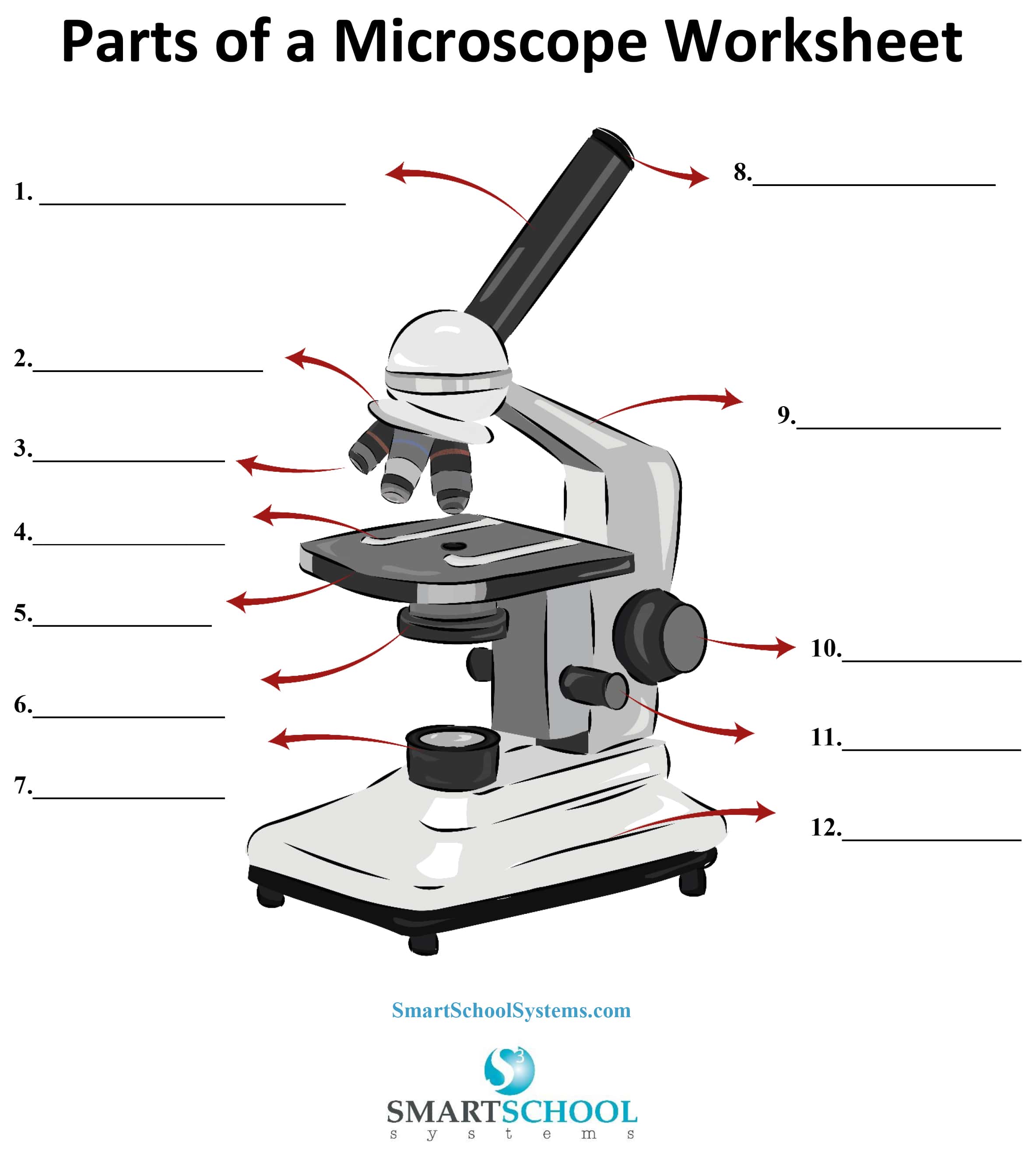
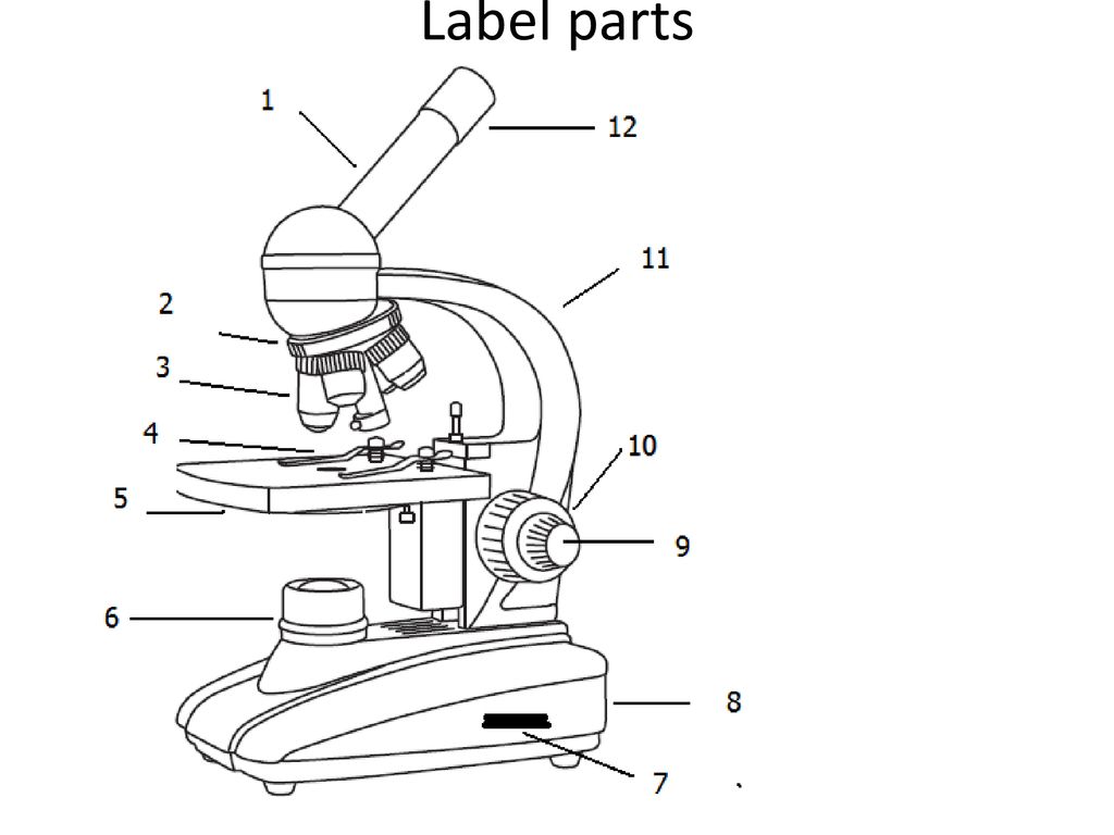
45 compound microscope diagram without labels
Structure of Cell: Definition, Types, Diagram, Functions - Embibe Cells in Latin means 'little rooms'. This name was given by Scientist Robert Hooke in the year \(1665\) who discovered the cell using a self-designed microscope. While studying a thin slice of cork (a substance that is obtained from the bark of a tree), Robert Hooke saw that the cork resembled the structure of a honeycomb consisting of many ... Simple cuboidal epithelium- structure, functions, examples Simple cuboidal epithelium definition. Simple cuboidal epithelium is a type of simple epithelium consisting of cube-shaped cells with rounds and more or less centrally located nucleus. They are mostly derived to suit the function of the particular organs better. Because the simple cuboidal epithelium has a single layer of cells, all the cube-shaped cells are directly attached to the basement ...
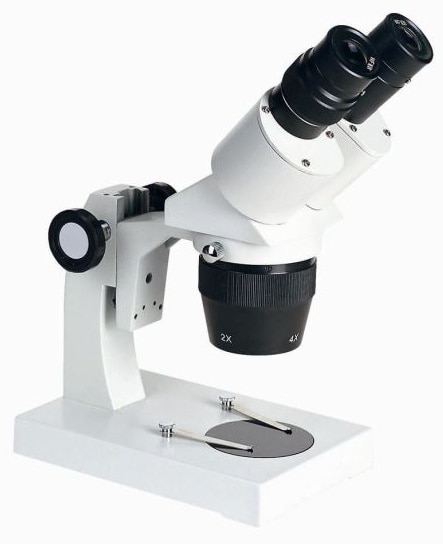
Binocular Microscope Anatomy - Parts and Functions with a Labeled Diagram Now, I will describe all these non-optical parts of the light compound microscope with the labeled diagrams. The body tube of the microscope. The body tube is the solid support for the optical and mechanical parts of the microscope. There are two basic types of stand in the body tube of a light compound microscope - upright stand and inverted ...
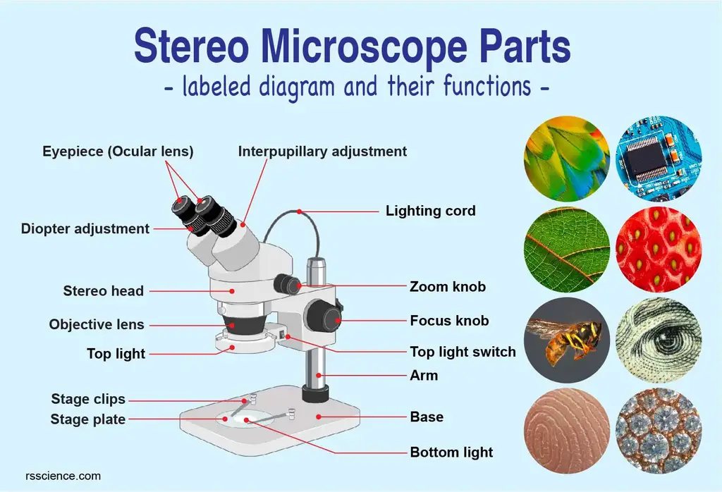
Compound microscope diagram without labels
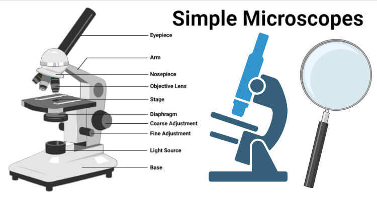
Parts of a Compound Microscope and Their Functions - NotesHippo Compound microscope mechanical parts (Microscope Diagram: 2) include base or foot, pillar, arm, inclination joint, stage, clips, diaphragm, body tube, nose piece, coarse adjustment knob and fine adjustment knob. Base: It's the horseshoe-shaped base structure of microscope. All of the other components of the compound microscope are supported ... Parts of a microscope with functions and labeled diagram - Microbe Notes Head - This is also known as the body. It carries the optical parts in the upper part of the microscope. Base - It acts as microscopes support. It also carries microscopic illuminators. Arms - This is the part connecting the base and to the head and the eyepiece tube to the base of the microscope. Course: s4: Biology , Topic: UNIT 3: MICROSCOPY - REB Care of the compound microscope The microscope is an expensive instrument that must be given proper care. Always ... amoeba, and paramecium. Observe, draw and label the visible parts under a light microscope. Avail these materials before you start: Petri-dishes, plate covers, pencil, transparent tape, microscope, agar powder, and permanent ...
Compound microscope diagram without labels. Introduction to the Compound Microscope: Parts & Uses Named for its pair of lenses and use of a light source, the compound light microscope was first used in the 17th century. The invention of this type of magnifier led to great discoveries of cells ... Scanning Electron Microscope (SEM) - Diagram, Working Principle ... Manfred von Ardenne developed the first version of the SEM in 1937. Q5. What is the cost of a scanning electron microscope? The price of a new electron microscope ranges between $80,000 to $10,000,000 and above depending on the customizations, configurations, resolution, components, and brand value. Light Microscope (Theory) - Amrita Vishwa Vidyapeetham Light microscope uses the properties of light to produce an enlarged image. It is the simplest type of microscope. Based on the simplicity of the microscope it may be categorized into: A) Simple microscope. B) Compound microscope. A) Simple microscope . It is uses only a single lens, e.g.: hand lens. Most of these are double convex or ... Compound Microscope- Definition, Labeled Diagram, Principle, Parts, Uses The optical microscope often referred to as the light microscope, is a type of microscope that uses visible light and a system of lenses to magnify images of small subjects. There are two basic types of optical microscopes: Simple microscopes. Compound microscopes. The term "compound" in compound microscopes refers to the microscope having ...
Histology, Staining - StatPearls - NCBI Bookshelf Medical Histology is the microscopic study of tissues and organs through sectioning, staining, and examining those sections under a microscope. Often called microscopic anatomy and histochemistry, histology allows for the visualization of tissue structure and characteristic changes the tissue may have undergone. Because of this, it is utilized in medical diagnosis, scientific study, autopsy ... how does a compound light microscope work - Shalon Devore A microscope will simply not work without proper lenses to begin with. ... A compound light microscope is a type of light microscope that uses a compound lens system meaning it operates through two sets of lenses to magnify the image. ... Microscope Diagram Labeled Unlabeled And Blank Parts Of A Microscope Science Printables Microscope Parts ... 15 Microscope Parts with Diagram, Location and Function - Study Read A compound microscope has about 15 parts that assist in viewing with a naked eye, a sample holder, a magnifying lens, and a light source. For the convenience of study, we can divide them based on their purpose in the instrument like. A) Parts that assist in viewing the object. B) Part that helps in the adjustment of lenses for a clear view. Plant Cells Vs. Animal Cells (With Diagrams) - Owlcation The vacuole has an important structural function, as well. When filled with water, the vacuole exerts internal pressure against the cell wall, which helps keep the cell rigid. A plant that is wilting has vacuoles that are no longer filled with water. While animal cells do not have a cell wall, chloroplasts, or a large vacuole, they do have one ...
Microscope Quiz: How Much You Know About Microscope Parts ... - ProProfs Projects light upwards through the diaphragm, the specimen, and the lenses. 5. Is used to regulates the amount of light on the specimen. Supports the slide being viewed. Moves the stage up and down for focusing. 6. Is used to support the microscope when carried. Moves the stage slightly to sharpen the image. Hair Under a Microscope - AnatomyLearner A compound microscope for examination purposes, ... Get more microscope hair-labeled diagrams on social media of anatomy learners. Cattle hair microscopic view ... You will find the imbricate scales in the cuticle of the horse hair without protrusion from the hair shaft. There are fine pigment granules distributed evenly in the medulla of the ... Stomach histology: Mucosa, glands and layers | Kenhub The innermost layer of the stomach wall is the gastric mucosa.It is formed by a layer of surface epithelium and an underlying lamina propria and muscularis mucosae. The surface epithelium is a simple columnar epithelium.It lines the inside of the stomach as surface mucous cells and forms numerous tiny invaginations, or gastric pits, which appear as millions of holes all throughout the stomach ... 20 most common lab equipment names, pictures and their uses 1. Microscope A light microscope placed on a white surface. Photo: pexels.com, @Artem Podrez Source: UGC. A microscope is a popular lab apparatus used to observe things that are too tiny to be observed by the naked human eye. There are many different types of microscopes.
How do electron microscopes work? - Explain that Stuff Scanning electron microscopes (SEMs) Most of the funky electron microscope images you see in books—things like wasps holding microchips in their mouths—are not made by TEMs but by scanning electron microscopes (SEMs), which are designed to make images of the surfaces of tiny objects. Just as in a TEM, the top of a SEM is a powerful electron gun that shoots an electron beam down at the ...
Chemical Laboratory Equipment Shapes and Usage | EdrawMax - Edrawsoft Step 3: To create a new lab apparatus drawing, go to [New] tab, find [Science and Education], and then click on [Laboratory Equipment]. This is where you'll find a wide range of templates to choose from and edit as needed. Step 4: To create a new one from zero, click on the "+" sign you can see in the picture above.
Simple Microscope - Parts, Functions, Diagram and Labelling Compound microscope - It comes with more than one lens and provides better magnification than the simple microscope. A compound microscope is also called a bright field microscope. It can provide magnification by up to 1,000 times. Stereo microscope/dissecting microscope - It can magnify objects by up to 300 times. It is used to visualize ...
Parts of a Microscope: Lesson for Kids - Study.com The light sits at the bottom of the microscope and directs light up through an image towards your eye. Now, look through the microscope like you would a pair of binoculars; these are the ocular ...
Pre Lab Video Coaching Activity Compound Microscope 45+ Pages Summary ... List all the parts of a compound microscope and give the function of each part. I also include labeled microscope diagrams for students to refer to at their lab tables along with a typed list of basic directions for using the microscope just in case students need a refresher of the basic microscope use information they explored in the online ...
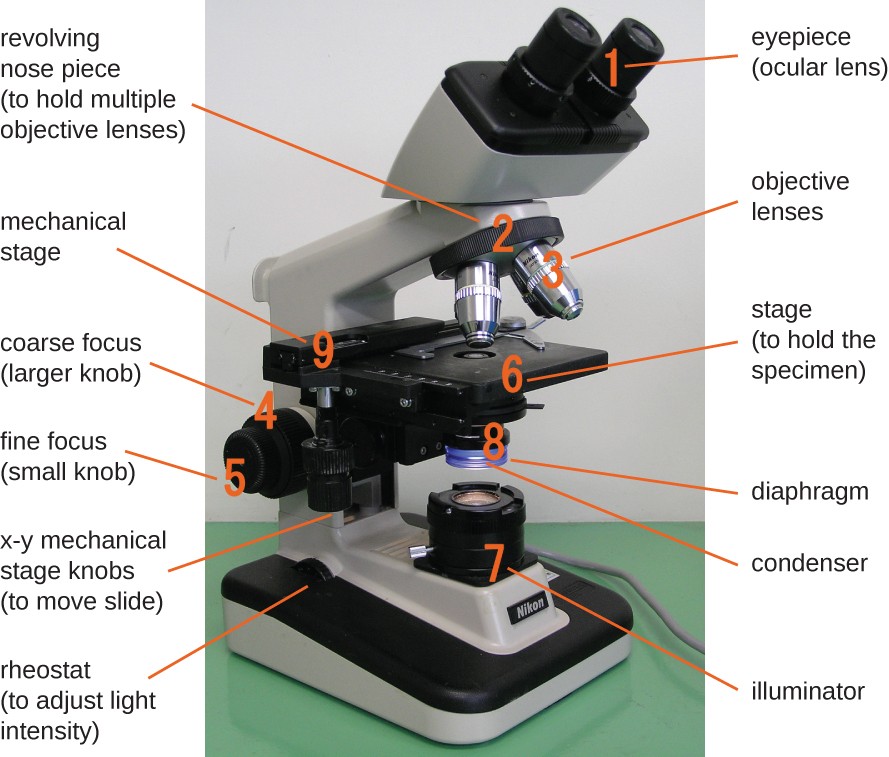
Microscope, Microscope Parts, Labeled Diagram, and Functions Revolving Nosepiece or Turret: Turret is the part of the microscope that holds two or multiple objective lenses and helps to rotate objective lenses and also helps to easily change power. Objective Lenses: Three are 3 or 4 objective lenses on a microscope. The objective lenses almost always consist of 4x, 10x, 40x and 100x powers. The most common eyepiece lens is 10x and when it coupled with ...
diagram shows a typical light microscope with its parts labeled ... diagram shows a typical light microscope with its parts labeled. Without a microscope, microorganisms would not be visible to the human eyes. _Ocular Lens (Eyepiece) Body Tube Revolving Nosepiece Objectives Arm Stage Clips Stage Coarse Adjustment Knob Fine Adjustment Knob Diaphragm Light Source Base...
Compound Microscope - Types, Parts, Diagram, Functions and Uses It comes with a wide body and base. Its distinct parts include a condenser, illumination, focus lock, mechanical stage, and a revolving nosepiece which can hold up to five objectives. It usually has a binocular head, which makes long-term observation easy. Image 22: An example of a research compound microscope.
(Get Answer) - Label the parts of the light microscope below using ... Explain why it does not belong. 3. light source, scanning power, objective lens, eyepiece. 4. compound light, transmission electron, light electron, scanning electron. 5. The ability of a microscope to show details clearly is called. 6. The ability of a microscope to increase an object's apparent sized is called. 7.
Euglena in microbiology movement, characteristics, and structure Euglena as a unicellular organism is too small to be seen with naked eyes, hence, in order to observe and study them, they are viewed under a compound microscope. These organisms divide their cell longitudinally for reproduction. They reproduce asexually through cell division by dividing down their length. Many species even produce dormant ...
How does a microscope work? - Explain that Stuff A compound microscope uses two or more lenses to produce a magnified image of an object, known as a specimen, placed on a slide (a piece of glass) at the base. The microscope rests securely on a stand on a table. Daylight from the room (or from a bright lamp) shines in at the bottom.
Course: s4: Biology , Topic: UNIT 3: MICROSCOPY - REB Care of the compound microscope The microscope is an expensive instrument that must be given proper care. Always ... amoeba, and paramecium. Observe, draw and label the visible parts under a light microscope. Avail these materials before you start: Petri-dishes, plate covers, pencil, transparent tape, microscope, agar powder, and permanent ...
Parts of a microscope with functions and labeled diagram - Microbe Notes Head - This is also known as the body. It carries the optical parts in the upper part of the microscope. Base - It acts as microscopes support. It also carries microscopic illuminators. Arms - This is the part connecting the base and to the head and the eyepiece tube to the base of the microscope.
Parts of a Compound Microscope and Their Functions - NotesHippo Compound microscope mechanical parts (Microscope Diagram: 2) include base or foot, pillar, arm, inclination joint, stage, clips, diaphragm, body tube, nose piece, coarse adjustment knob and fine adjustment knob. Base: It's the horseshoe-shaped base structure of microscope. All of the other components of the compound microscope are supported ...
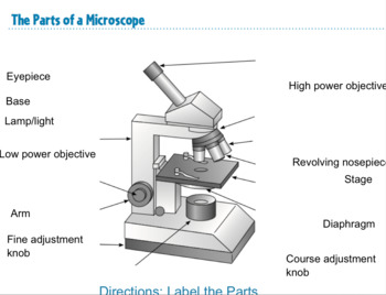
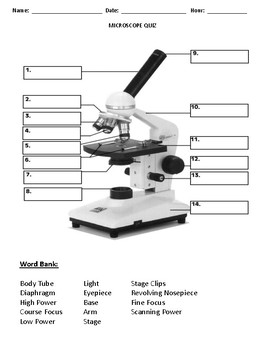

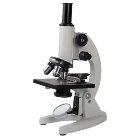















Post a Comment for "45 compound microscope diagram without labels"