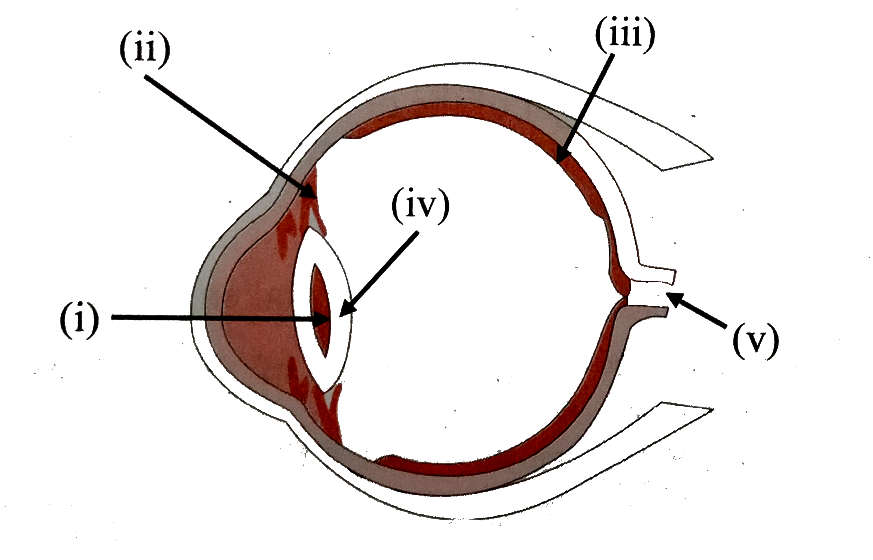
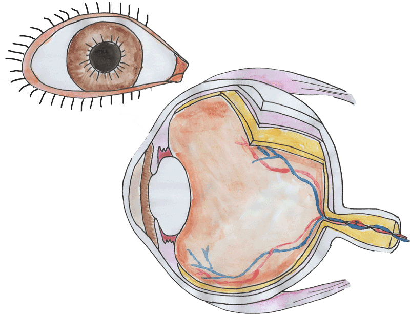
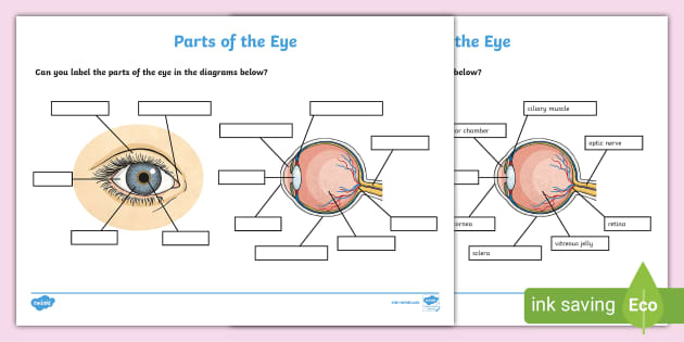
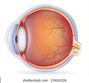
38 the human eye without labels
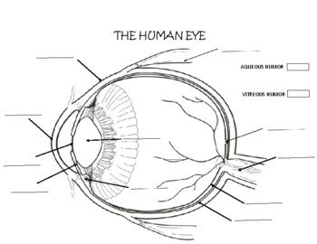
Anatomy of the eye: Quizzes and diagrams | Kenhub Here you can see all of the main structures in this area. Spend some time reviewing the name and location of each one, then try to label the eye yourself - without peeking! - using the eye diagram (blank) below. Unlabeled diagram of the eye Click below to download our free unlabeled diagram of the eye. Anatomy of the Human Eye - News-Medical.net The light passing through cornea, pupil, and lens gets focused on the retinal membrane. In addition to tissue components, retina is made up of two types of cells: rod cells and cone cells. The ...
File:Diagram of human eye without labels.svg - Wikimedia Size of this PNG preview of this SVG file: 410 × 430 pixels. Other resolutions: 229 × 240 pixels | 458 × 480 pixels | 732 × 768 pixels | 976 × 1,024 pixels | 1,953 × 2,048 pixels. Original file (SVG file, nominally 410 × 430 pixels, file size: 277 KB) File information. Structured data.
The human eye without labels
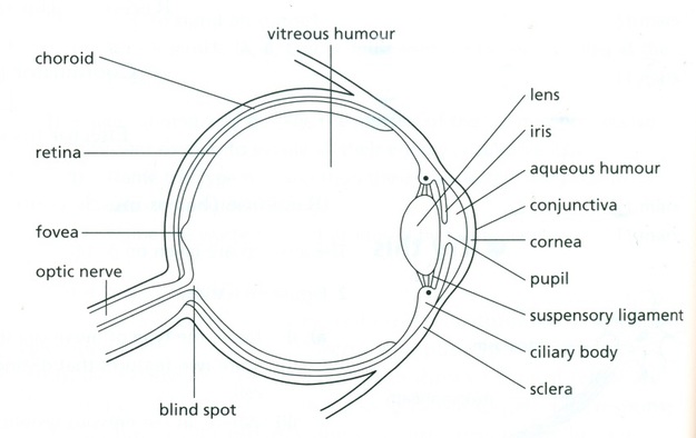
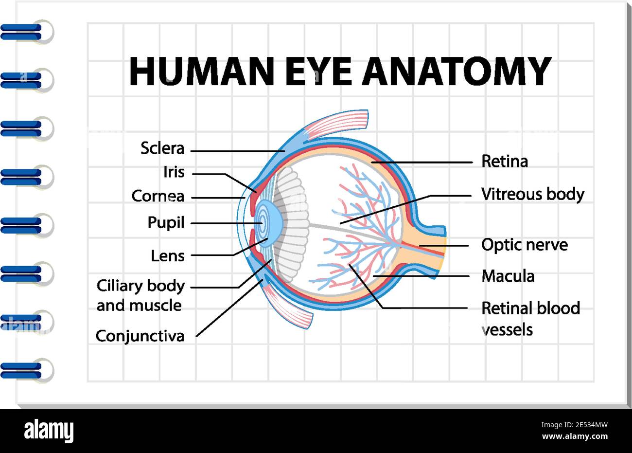
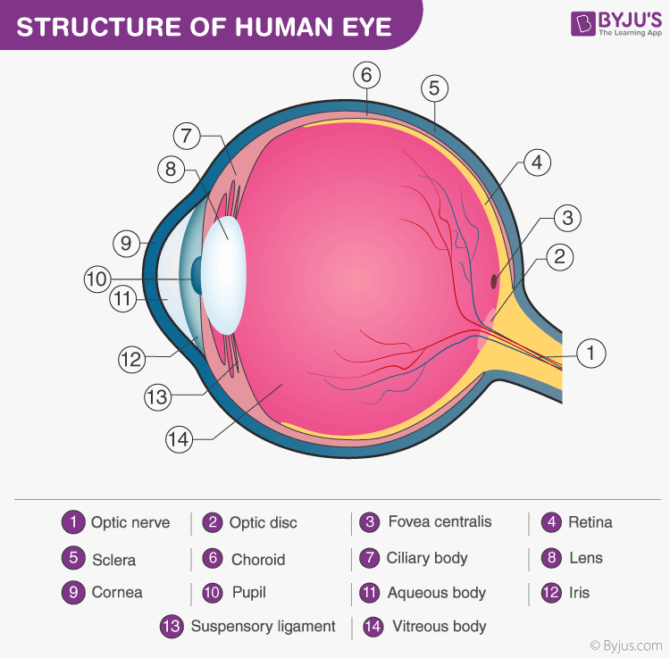
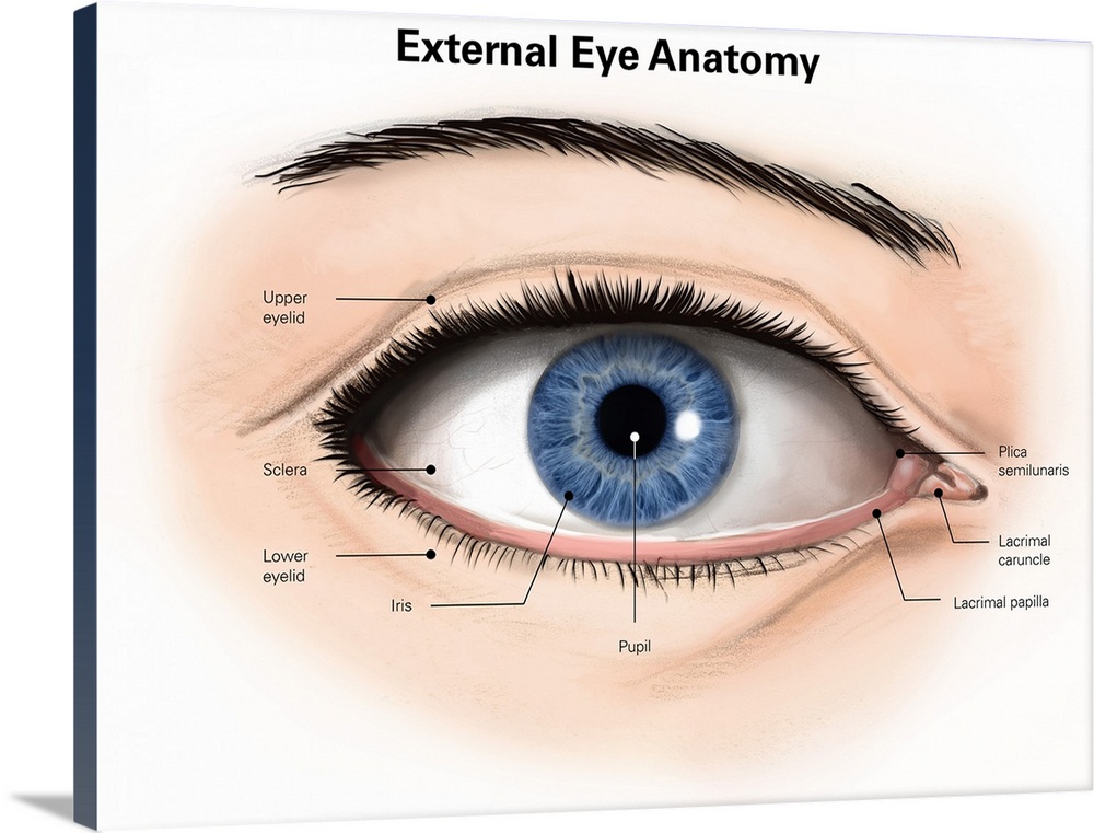
Eye Anatomy: 16 Parts of the Eye & Their Functions - Vision Center The following are parts of the human eyes and their functions: 1. Conjunctiva. The conjunctiva is the membrane covering the sclera (white portion of your eye). The conjunctiva also covers the interior of your eyelids. Conjunctivitis, often known as pink eye, occurs when this thin membrane becomes inflamed or swollen. Human Eye - Definition, Structure, Function, Parts, Diagram - BYJUS Structure of Human Eye. A human eye is roughly 2.3 cm in diameter and is almost a spherical ball filled with some fluid. It consists of the following parts: Sclera: It is the outer covering, a protective tough white layer called the sclera (white part of the eye). Cornea: The front transparent part of the sclera is called the cornea. human eye | Definition, Anatomy, Diagram, Function, & Facts The protrusion of the eyeballs—proptosis—in exophthalmic goitre is caused by the collection of fluid in the orbital fatty tissue. The eyelids eyelid It is vitally important that the front surface of the eyeball, the cornea, remain moist.
The human eye without labels. Common Types of Warehouse Labels - ID Label Inc. Warehouse Rack Beam Labels. The most common type of warehouse label is found on rack beams. These labels mark each rack bay location and are used to identify products for storing, picking and inventory management. They typically include a one- or two-dimensional barcode image and human-readable letters and numbers. Structure Of Human Eye Without Label - NicePNG Structure Of Human Eye Without Label is a totally free PNG image with transparent background and its resolution is 952x698. You can always download and ... Eye Diagram With Labels and detailed description - BYJUS Iris is the coloured part of the eye and controls the amount of light entering the eye by regulating the size of the pupil. The lens is located just behind the iris. Its function is to focus the light on the retina. The optic nerve transmits electrical signals from the retina to the brain. Pupil is the opening at the centre of the iris. Label Parts of the Human Eye - University of Dayton Parts of the Eye. Select the correct label for each part of the eye. The image is taken from above the left eye. Click on the Score button to see how you did. Incorrect answers will be marked in red. ...
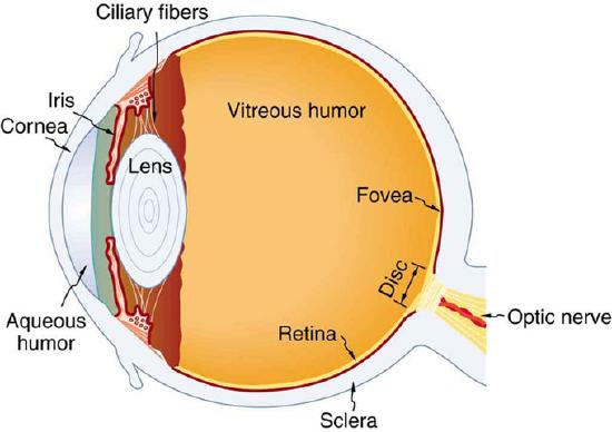
label the eye worksheet eye diagram human worksheet eyeball learning layers anatomy without parts labels worksheets eyes science clipart structure grade body exercise structures The Best Free Eye Drawing Images. Download From 4424 Free Drawings Of getdrawings.com labeled purposegames unlabelled labeling quizlet healthiack getdrawings einzigartig studyblue detailed Structure and Function of the Human Eye - ThoughtCo The main parts of the human eye are the cornea, iris, pupil, aqueous humor, lens, vitreous humor, retina, and optic nerve. Light enters the eye by passing through the transparent cornea and aqueous humor. The iris controls the size of the pupil, which is the opening that allows light to enter the lens. Light is focused by the lens and goes ... PDF Eye Anatomy Handout - National Institutes of Health of light entering the eye. Lens: The lens is a clear part of the eye behind the iris that helps to focus light, or an image, on the retina. Macula: The macula is the small, sensitive area of the retina that gives central vision. It is located in the center of the retina. Optic nerve: The optic nerve is the largest sensory nerve of the eye. 60,892 Human eye anatomy Images, Stock Photos & Vectors - Shutterstock Find Human eye anatomy stock images in HD and millions of other royalty-free stock photos, illustrations and vectors in the Shutterstock collection. Thousands of new, high-quality pictures added every day.
Category:Human eyes - Wikimedia Commons Black eyes by megamoto85 (cropped).jpg 925 × 673; 148 KB Blue Eyed Girl - Flickr - rcstanley.jpg 529 × 622; 49 KB Blue-Green Eye miosis.jpg 917 × 688; 451 KB label the ear worksheet Picture Front Of The Eye Without Labels Clipart 20 Free Cliparts clipground.com eye human diagram worksheet eyeball learning layers without anatomy labels parts eyes worksheets clipart science structure grade clipground body structures 14 Best Images Of Ear Hearing Worksheets - Listening Ear Craft Template Structure of the Human Eye - Health Jade The eye is a hollow, spherical structure about 2.5 centimeters in diameter. Its wall has three distinct layers—an outer (fibrous) layer, a middle (vascular) layer, and an inner (nervous) layer. The spaces within the eye are filled with fluids that help maintain its shape. Figure 6. Structure of the human eye. The Eyes (Human Anatomy): Diagram, Optic Nerve, Iris, Cornea ... - WebMD The weaker eye, which may or may not wander, is called the "lazy eye." Astigmatism: A problem with the curve of your cornea. If you have it, your eye can't focus light onto the retina the way it...
Eye Anatomy: A Closer Look At the Parts of the Eye - All About Vision In a number of ways, the human eye works much like a digital camera: Light is focused primarily by the cornea — the clear front surface of the eye, which acts like a camera lens. The iris of the eye functions like the diaphragm of a camera, controlling the amount of light reaching the back of the eye by automatically adjusting the size of the ...
Human eye - Wikipedia The human eye is a sensory organ, part of the sensory nervous system, that reacts to visible light and allows us to use visual information for various purposes including seeing things, keeping our balance, and maintaining circadian rhythm . The eye can be considered as a living optical device.
The Human Eye | Boundless Physics | | Course Hero The human eye is the gateway to one of our five senses. The human eye is an organ that reacts with light. It allows light perception, color vision and depth perception. A normal human eye can see about 10 million different colors! There are many parts of a human eye, and that is what we are going to cover in this atom. Properties
What Does the Eye Look Like? - Diagram of the Eye | Harvard Eye Associates Vitreous Gel: A thick, transparent liquid that fills the center of the eye. It is mostly water and gives the eye its form and shape. Our eyes are vital for seeing the world around us. Keep them healthy by maintaining regular vision exams. Contact Harvard Eye Associates at 949-951-2020 or harvardeye.com to schedule an appointment today.
Eye Anatomy: Parts of the Eye and How We See Behind the anterior chamber is the eye's iris (the colored part of the eye) and the dark hole in the middle called the pupil. Muscles in the iris dilate (widen) or constrict (narrow) the pupil to control the amount of light reaching the back of the eye. Directly behind the pupil sits the lens. The lens focuses light toward the back of the eye.
Quiz: Label The Parts Of The Eye - ProProfs Quiz Quiz: Label The Parts Of The Eye. Do you know the anatomy of the human eye very well? Can you label the parts of the eye in the quiz below? Give it a try and evaluate yourself. The eye has many important parts, each with different functions, including the cornea, pupil, sclera, and many more. Can you tell where these parts are located and what ...
How Do We See Light? | Ask A Biologist - Arizona State University The human eye has over 100 million rod cells. Cones require a lot more light and they are used to see color. We have three types of cones: blue, green, and red. The human eye only has about 6 million cones. Many of these are packed into the fovea, a small pit in the back of the eye that helps with the sharpness or detail of images.
Eye Diagram Teaching Resources | Teachers Pay Teachers The Human Eye Overview Reading Comprehension and Diagram Worksheet. by. Teaching to the Middle. 4.7. (65) $1.50. Zip. This passage briefly describes the human eye (900-1000 Lexile). 14 questions (matching and multiple choice) assess students' understanding. Students label a diagram of 6 parts of the eye.
The Human Eye - Diagram, Parts, Working, Function and Work of The Lens Sclera: The sclera is the protective outer layer, a strong white coating that protects the eyes (white part of the eye). Cornea: The cornea is the sclera's translucent front part. The cornea allows light to flow through and into the eye. Iris: The iris is a black muscular tissue and ring-like structure behind the cornea. The eye's colour is determined by the colour of the iris.
File:Diagram of human eye without labels.svg - Wikipedia File:Diagram of human eye without labels.svg. Size of this PNG preview of this SVG file: 410 × 430 pixels. Other resolutions: 229 × 240 pixels | 458 × 480 ...
Diagram of the Eye Side View No Labels Illustration - Twinkl Sep 18, 2019 - Diagram of the Eye Side View No Labels,Eye,Science,Human Body,Biology,Diagram,Human,Pupil,Sense,Organ,Sclera,Cornea,Iris,Lens,Retina,Optic ...
Placeholders in Form Fields Are Harmful - Nielsen Norman Group May 11, 2014 · Labels and Placeholders. Labels tell users what information belongs in a given form field and are usually positioned outside the form field. Placeholder text, located inside a form field, is an additional hint, description, or example of the information required for a particular field. These hints typically disappear when the user types in the ...
How the Eyes Work | National Eye Institute - National Institutes of Health How the Eyes Work. All the different parts of your eyes work together to help you see. First, light passes through the cornea (the clear front layer of the eye). The cornea is shaped like a dome and bends light to help the eye focus. Some of this light enters the eye through an opening called the pupil (PYOO-pul).
Label Parts of the Human Ear - University of Dayton Parts of the Ear. Select the correct label for each part of the ear. Click on the Score button to see how you did. Incorrect answers will be marked in red.
Human Eye Explorer • Work without program installation. With Human Eye Explorer you can: EVERYTHING IN MOTION. ... Label, cut, color, isolate and set transparencies. EXCITING PRESENTATIONS. Change perspectives, ... The virtual 3D model of the human eye is the central element of the application. It contains all important structures like the eyeball and its ...
label muscles worksheet Picture Front Of The Eye Without Labels Clipart - Clipground clipground.com. eye human diagram worksheet layers eyeball learning anatomy labels without clipart eyes parts worksheets science structure clipground grade body structures. Posterior View Of The Superficial Muscles Of The Arm | ClipArt ETC etc.usf.edu
human eye | Definition, Anatomy, Diagram, Function, & Facts The protrusion of the eyeballs—proptosis—in exophthalmic goitre is caused by the collection of fluid in the orbital fatty tissue. The eyelids eyelid It is vitally important that the front surface of the eyeball, the cornea, remain moist.
Human Eye - Definition, Structure, Function, Parts, Diagram - BYJUS Structure of Human Eye. A human eye is roughly 2.3 cm in diameter and is almost a spherical ball filled with some fluid. It consists of the following parts: Sclera: It is the outer covering, a protective tough white layer called the sclera (white part of the eye). Cornea: The front transparent part of the sclera is called the cornea.
Eye Anatomy: 16 Parts of the Eye & Their Functions - Vision Center The following are parts of the human eyes and their functions: 1. Conjunctiva. The conjunctiva is the membrane covering the sclera (white portion of your eye). The conjunctiva also covers the interior of your eyelids. Conjunctivitis, often known as pink eye, occurs when this thin membrane becomes inflamed or swollen.











Post a Comment for "38 the human eye without labels"