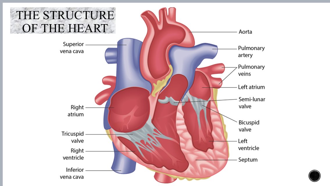
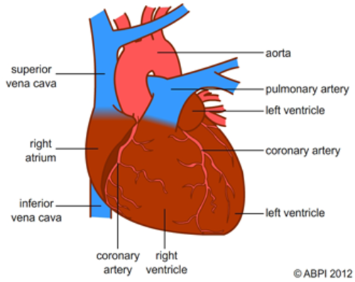
38 heart structure with labels
Simple heart diagram | Simple heart diagram labeled - Pinterest Internal structure of human heart shows four chambers viz. two atria and two ventricles and couple of blood vessels opening into them. The wall of two ventricles are strong and sturdy when compared to atria. Before we start, we shall recall the basic proportions of heart and its chambers. The right Auricle is larger than left. Human Heart: Label the diagram 1 - Liveworksheets Human Heart: Label the diagram 1 worksheet. Live worksheets > English. Human Heart: Label the diagram 1. Study the figure carefully.Label the 10 parts of the human heart A-J. ID: 1781041. Language: English. School subject: Biology. Grade/level: 9-12. Age: 14+.
Heart Diagram for Kids - Bodytomy As you can see in the diagram of the heart, that heart is divided in four chambers, namely, right atrium, left atrium, right ventricle and left ventricle. Each of the chambers is separated by a muscle wall known as Septum. The left side of the heart receives oxygen rich blood from the lungs and pumps it out the whole body.
Heart structure with labels
Label the heart - Teaching resources - Wordwall 10000+ results for 'label the heart'. Label the Heart Labelled diagram. by Banksm. Cardiovascular system - Label the heart Labelled diagram. by Temorris. KS4 PE. Label the Heart diagram (L3) Labelled diagram. by Jenniferross. Y9 Biology. Heart Diagram with Labels and Detailed Explanation - BYJUS Diagram of Heart. The human heart is the most crucial organ of the human body. It pumps blood from the heart to different parts of the body and back to the heart. The most common heart attack symptoms or warning signs are chest pain, breathlessness, nausea, sweating etc. The diagram of heart is beneficial for Class 10 and 12 and is frequently ... Human Heart (Anatomy): Diagram, Function, Chambers, Location in ... - WebMD Human Heart (Anatomy): Diagram, Function, Chambers, Location in Body The right atrium receives blood from the veins and pumps it to the right ventricle. The right ventricle receives blood from the...
Heart structure with labels. Label Heart Anatomy Diagram Printout - EnchantedLearning.com | Heart ... Feb 7, 2012 - Label Heart Interior Anatomy Diagram Printout. Feb 7, 2012 - Label Heart Interior Anatomy Diagram Printout. Pinterest. Today. Explore. ... Heart Structure. Science Diagrams. Download for free Blank Heart Diagram #1664321, download othes parts of the heart to label for free. Megan Ensign. Nursing School. heart diagram and labels The Anatomy and Physiology of Animals/Circulatory System Worksheet. 11 Images about The Anatomy and Physiology of Animals/Circulatory System Worksheet : walls label label beginning Heart Diagram With Labels And Blood Flow, labelled diagram of heart a level - Clip Art Library and also labelled diagram of heart a level - Clip Art Library. Heart: Anatomy and Function - Cleveland Clinic Heart. Your heart is the main organ of your cardiovascular system, a network of blood vessels that pumps blood throughout your body. It also works with other body systems to control your heart rate and blood pressure. Your family history, personal health history and lifestyle all affect how well your heart works. Appointments 800.659.7822. Label the Heart Diagram | Quizlet Label the Heart STUDY Learn Write Test PLAY Match Created by bluesas9 Terms in this set (15) Superior Vena Cava ... Right Ventricle ... Left Atrium ... Atrioventricular/Tricuspid Valve ... Atrioventricular/Mitral Valve ... Septum ... Right Atrium ... Semi-lunar Valves ... Left Pulmonary Veins ... Right Pulmonary Veins ... Left Pulmonary Arteries
Human Heart - Anatomy, Functions and Facts about Heart The heart wall is made up of 3 layers, namely: Epicardium - Epicardium is the outermost layer of the heart. It is composed of a thin-layered membrane that serves to lubricate and protect the outer section. Myocardium - This is a layer of muscle tissue and it constitutes the middle layer wall of the heart. Structure and Function of the Heart - Medical News The heart is a muscle whose working mechanism is made possible by the many parts that operate together. The organ is divided into several chambers that take in and distribute oxygen-poor or oxygen ... diagram of heart labelled liver cell drawing diagram draw label ultrastructure paintingvalley animal example labelling diagrams ze kig source. Heart Models . heart anatomy models chambers biologycorner labeled internal label vessels 3d pulmonary cardiac publisher follow google circulatory. Label The Heart Worksheets (SB6634) - SparkleBox www ... Structure of Heart (With Diagram) | Circulatory System | Human Physiology The heart is consisting of three layers: 1. Pericardium or outer covering layer: The heart lies in a double membranous sac of pericardium with serous fluid between the two layers. This is known as pericardial fluid. By its lubricating action, the heart can move freely or contracts and expands without any injury.
Heart Labeling Quiz: How Much You Know About Heart Labeling? Here is a Heart labeling quiz for you. The human heart is a vital organ for every human. The more healthy your heart is, the longer the chances you have of surviving, so you better take care of it. Take the following quiz to know how much you know about your heart. Questions and Answers 1. What is #1? 2. What is #2? 3. What is #3? 4. What is #4? Diagrams, quizzes and worksheets of the heart | Kenhub Worksheet showing unlabelled heart diagrams. Using our unlabeled heart diagrams, you can challenge yourself to identify the individual parts of the heart as indicated by the arrows and fill-in-the-blank spaces. This exercise will help you to identify your weak spots, so you'll know which heart structures you need to spend more time studying ... Human Heart (Anatomy): Diagram, Function, Chambers, Location in ... - WebMD Human Heart (Anatomy): Diagram, Function, Chambers, Location in Body The right atrium receives blood from the veins and pumps it to the right ventricle. The right ventricle receives blood from the... Heart Diagram with Labels and Detailed Explanation - BYJUS Diagram of Heart. The human heart is the most crucial organ of the human body. It pumps blood from the heart to different parts of the body and back to the heart. The most common heart attack symptoms or warning signs are chest pain, breathlessness, nausea, sweating etc. The diagram of heart is beneficial for Class 10 and 12 and is frequently ...
Label the heart - Teaching resources - Wordwall 10000+ results for 'label the heart'. Label the Heart Labelled diagram. by Banksm. Cardiovascular system - Label the heart Labelled diagram. by Temorris. KS4 PE. Label the Heart diagram (L3) Labelled diagram. by Jenniferross. Y9 Biology.

Limehurst Academy PE on Twitter: "GCSE PE Revision: cardiovascular system structure of heart and ...


Post a Comment for "38 heart structure with labels"