41 stage diagram with labels
A Labelled Diagram Of Meiosis with Detailed Explanation Well-Labelled Diagram for Meiosis Meiosis I Prophase I Here, the chromosomes begin to condense. Prophase I is divided into five different stages: Leptotene Zygotene Pachytene Diplotene Diakinesis Metaphase I The homologous pairs of chromosomes are aligned on the equatorial plate. Anaphase I KS2 Label the skeleton - Teaching resources Label The Bones of the Skeleton 7.12 8.12 Labelled diagram. by Cdunsdon. Label The Bones of the Skeleton - AQA GCSE Labelled diagram. by Cdemosthenous. Label a Human Skeleton Labelled diagram. by Harrisond. KS1 KS2 Y2 Y3 Y4 Y5 Y6 Science. The Skeleton and the Muscles Quiz. by Afiahntiriakuff.
Solved Glycolysis, Krebs, or electron transport Drag each ... Glycolysis, Krebs, or electron transport Drag each of the descriptions on the left to the stage or stages of cellular respiration during which it is found. If the description could be classified in more than one stage, place it in the intersecting parts of those circles of this Venn diagram. Two labels regarding NADH have been filled in for you.

Stage diagram with labels
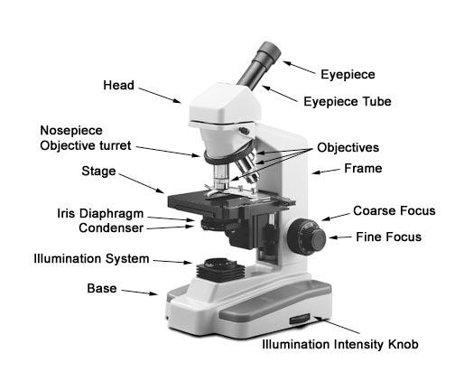
Microscope Parts and Functions With Labeled Diagram and ... Most specimens are mounted on slides, flat rectangles of thin glass. The specimen is placed on the glass and a cover slip is placed over the specimen. This allows the slide to be easily inserted or removed from the microscope. It also allows the specimen to be labeled, transported, and stored without damage. Parts of Microscope, Function, Names & Labeled Diagram ... Microscope parts labeled diagram gives us all the information about its parts and their position in the microscope. Microscope Parts Labeled Diagram The principle of the Microscope gives you an exact reason to use it. It works on the 3 principles. Magnification Resolving Power Numerical Aperture. Parts of Microscope Head Base Arm Eyepiece Lens Stages Of Meiosis Diagram Labeled - Wiring Diagrams Stages Of Meiosis Diagram Labeled Our chromosome number is For the existence of any species chromosome number should be maintained. We inherited the genetic program from our parents. Meiosis is the process in eukaryotic, sexually-reproducing animals that reduces In the diagram below, the red chromosomes are the ones inherited from the.
Stage diagram with labels. Mitosis: Phases, Diagram, Stage, and Checkpoints Diagram of Mitosis Phases Stages of Mitosis i. Prophase o Condensation of chromatin into chromosomes occurs. o Centrioles move to opposite ends of the cells. o First the nucleolus and then the nucleus disappear. Prophase Diagrams - Wiring Diagrams Free Prophase, metaphase, anaphase, and telophase. Diagrams of Mitosis - the process of cell division via mitosis occurs in a series of stages including prophase, metaphase, Anaphase and Telophase. It is easy to describe the stages of mitosis in the form of diagrams showing the dividing cell (s) at each of the main stages of the process. Diagram - Key Stage Wiki Diagram Key Stage 1 Meaning A diagram is a picture with labels. Key Stage 2 Meaning A diagram is a picture with labels or a picture used to help explain something. About Diagrams Diagrams can be used to identify parts of an object. Diagrams can be used to explain a process without lots of writing. Examples Key Stage 3 Meaning Volvox Diagram Labeled - Wiring Diagrams You should make a label that represents your brand and creativity, at the same time you shouldn't forget. Related: euglena diagram labeled, earthworm diagram labeled, human teeth diagram labeled, kidney diagram labeled, labelled diagram of chloroplast, human heart diagram labeled for kids, volvox slide labeled, skin diagram with labels, human ...
Compound Microscope Parts - Labeled Diagram and their ... The stage is a flat platform that supports the slides. The stage has an opening (called aperture) for the illuminating beam of light to pass through. The stage clips hold the slides in place. If your microscope has a mechanical stage, the slide secured on the slide holder can be moved in two perpendiculars (X - Y) directions by turning two ... Stage Gate Process | Download | Easy to Edit | PowerSlides™ The Stage Gate process model focuses on the innovation process and is also referred to as the waterfall process. It is a project management technique, in which an initiative or project takes place, divided over several stages. The stages technique breaks new product development projects into five phases - scoping, business case creation, development, testing … Continue reading "Stage Gate ... Solved 5' 96. A eukaryotic translation complex during the ... A eukaryotic translation complex during the elongation stage is represented by the diagram above. Which of the following labels on the diagram is NOT correct? (A) Location of the mRNA cap structure (B) Direction of ribosome movement (C) Anticodon of a tRNA molecule (D) 5' end of a tRNA molecule (E) Carboxyl-terminus of the growing peptide chain You Parts of the Stage - Diagram and Quiz/Worksheet | Teaching ... Description A labeled diagram of a proscenium stage, as well as an identical fill-in-the-blank worksheet. Use in introductory theatre or stagecraft classes as a quiz or bell-ringer. Teachers Pay Teachers 1M followers More information Parts of the Stage - Diagram and Worksheet Find this Pin and more on You Will Be Schooled Here by Laura Goldstein.
Parts of a microscope with functions and labeled diagram The coarse adjustment knob moves the stage up and down to bring the specimen into focus. The fine adjustment knob brings the specimen into sharp focus under low power and is used for all focusing when using high-power lenses. Q. List down the 18 parts of a Microscope. 1. Ocular Lens (Eye Piece) 2. Diopter Adjustment 3. Head 4. Nose Piece 5. Parts of the Stage - Diagram and Quiz/Worksheet by Laura ... Description A labeled diagram of a proscenium stage, as well as an identical fill-in-the-blank worksheet. Use in introductory theatre or stagecraft classes as a quiz or bell-ringer. Total Pages 2 pages Answer Key Included Teaching Duration N/A Report this Resource to TpT Reported resources will be reviewed by our team. Microscope, Microscope Parts, Labeled Diagram, and Functions The description given below summarize the brief description of microscope parts used to visualize the microscopic specimens such as animal cells, plant cells, microbes, bacteria, viruses, microorganisms etc. The Microscopes parts divided into three different structural parts Head, Base, and Arms. Head/Body: It contain the optical parts in the ... Mastering Biology CH 13 homework Flashcards | Quizlet Drag the labels to fill in the targets beneath each diagram of a cell. Note that the diagrams are in no particular order.Drag labels of Group 1 to identify the stage of meiosis depicted in each diagram.Drag labels of Group 2 to identify whether the configuration of the chromosomes related to crossing over is possible or not.
Print Microbiology Lab 2 (Microscopy, Gram stain, intro to enterotube II) flashcards | Easy ...
Tecrider.com - Your free online stage plot designer Design and share your Stage Plot online in a simple way just by drag & drop your instruments on the stage. We help you to create your Technical Rider online to have a much easier show experience. Once you've created your Tech Rider online, you can share it with your band members, download your Stage Plot as PDF or send it out to your venues.

Lauren's Blog: October 2010
Human Ear Diagram - Bodytomy Human Ear Diagram. Wondering what is the structure of the human ear, and how it performs the function of hearing? Look no further, this Bodytomy article gives you a labeled human ear diagram and also explains the functions of its different components.
Design Chevron and Circular Diagrams
Spirogyra Labelled Diagram Spirogyra (common names include water silk, mermaid's tresses, and blanket weed) is a genus of filamentous charophyte green algae of the order Zygnematales, named for the helical or spiral arrangement of the chloroplasts that is characteristic of the genus. Draw a labelled diagram of Spirogyra. 51 Differentiate between flying lizard and bird.
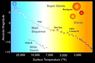
Hertzsprung-Russell Diagram
Draw a labeled diagram to show the metaphase stage of ... Given below is a diagram representing a stage during mitotic cell division in an animal cell. Examine it carefully and answer the questions which follow. (a) Identify the stage. Give one reason in support of your answer. (b) Name the cell organelle that forms the 'aster'. (c) Name the parts labelled 1, 2 and 3.
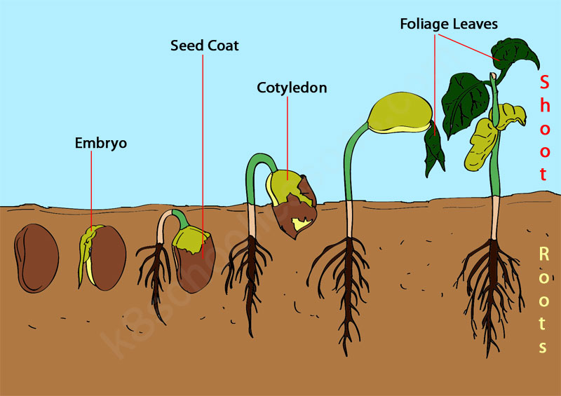
Seed Germination Quiz | Science Lessons for kids | Science Quiz
DOC Mitosis: Labeled Diagram Mitosis: Labeled Diagram Mitosis is a process of cell division which results in the production of two daughter cells from a single parent cell. The daughter cells are identical to one another and to the original parent cell.
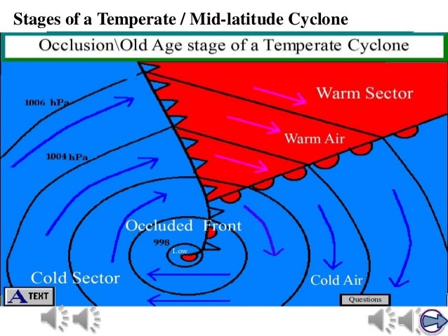
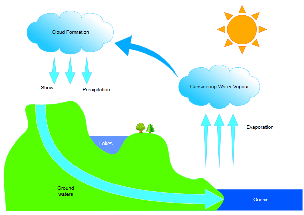
Temperate Cyclones
Mastering Biology CH 13 homework Flashcards - Quizlet Complete the diagram to show the life cycle of a typical animal. Follow these steps: Drag labels of Group 1 to identify each stage of the life cycle. Drag labels of Group 2 to identify the ploidy level at each stage. Drag labels of Group 3 to identify the process by which each stage occurs. Labels can be used once, more than once, or not at all.

Ophthalmology | Ophthalmologist: PTERYGIUM
Diagram of a Theater - Greek Theatre Notes V. Diagram of Theater A. theatron - where the audience sits B. orchestra - the stage; where the actors performed C. thymele - altar to Dionysus in center of orchestra D. skene - building used as a dressing room E. proskeneion - façade of skene building used as a backdrop F. parados - entrance & exit to the theater used by actors & chorus
Post a Comment for "41 stage diagram with labels"