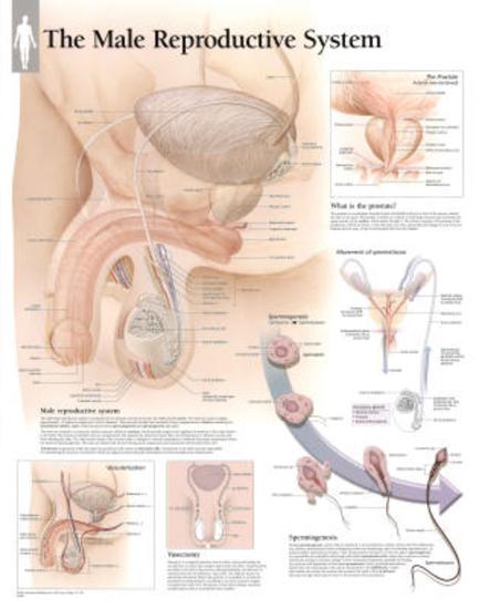
39 male reproductive system diagram without labels
Male reproductive organs: Anatomy and function - Kenhub The testes (singular: testis) are two oval-shaped male internal genital organs found within the scrotum. Their function is to produce sperm and the hormone testosterone. Testicular diagram with neighbouring structures Testes comprise an intricate network of tubules and dispersed secretory cells. nucellus is haploid or diploid Answer: Yes, if the embryo develops from the cells of nucellus or integument, it will be diploid. So they both differ in haploidy. Thus diploid number of chromosomes is present. T
Egg cell - Wikipedia The egg cell, or ovum(plural ova), is the female reproductivecell, or gamete, in most anisogamousorganisms (organisms that reproduce sexuallywith a larger, female gamete and a smaller, male one). The term is used when the female gamete is not capable of movement (non-motile).
Male reproductive system diagram without labels
Digestive System Answers Quizlet Worksheet Mouth: Human mouth consists of two parts There is a PDF and an editable version of the worksheets Reproductive system b 9 org are unblocked Below is a labeled diagram to help the students correctly label the organs and a functions worksheet to help write in the functions of each organ Below is a labeled diagram to help the students correctly ... Female reproductive organs: Anatomy and functions | Kenhub Our labeled diagrams and quizzes on the female reproductive system are the best place to start. The uterus is supplied mainly by the uterine artery which arises from the internal iliac artery. The superior branch of the uterine artery supplies the body and fundus, while the inferior branch supplies the cervix. Organs on Left Side of Body: Inside and Out, From Head to Toe Cones and rods The eye contains about 6 million cone cells and 90 million rod cells. Left lung Your left lung has only two lobes while your right lung has three lobes. This asymmetry allows room...
Male reproductive system diagram without labels. Male Reproductive System: Functions, Organs & Anatomy ... Diagram of the male reproductive system So what are these structures? Well, first and foremost we have the testes, or testis (singular). The testes are paired structures in the male whose function... 11 Major Reproductive System Diseases in Women | New ... The reproductive system in women consists of many parts such as the ovaries, fallopian tubes, vagina, cervix, external genitals and uterus, but these different parts are also susceptible to many diseases that can harm fertility or lead to further serious illness. Bio 101 ch.29 Flashcards | Quizlet "start the life cycle with the mature sporophyte stage in target 1. Not all labels will be used a) haploid gametes undergo fertilization, forming a diploid zygote. b) Haploid eggs form in archegonia, and haploid sperm form in antherida c) Haploid gametes undergo meiosis, forming a … Meiosis- definition, purpose, stages, applications with ... Meiosis definition. Meiosis is a type of cell division in sexually reproducing eukaryotes, resulting in four daughter cells (gametes), each of which has half the number of chromosomes as compared to the original diploid parent cell. The haploid cells become gametes, which by union with another haploid cell during fertilization defines sexual ...
Anatomy of the male canine abdomen and pelvis on ... - IMAIOS CT images are from a healthy 6-year-old castrated male dog. In this module of the animal atlas vet-Anatomy is displayed the cross-sectional labeled anatomy of the canine abdominal cavity and the pelvis on a Computed Tomography (CT) and on 3D images of the abdomen of the dog. CT images are available in 3 different planes (transverse, sagittal ... Understanding Intersectionality. The concept of ... - Medium 12/10/2017 · The concept of Intersectionality was introduced by Kimberle Crenshaw in an article in 1989. It refers to the overlapping or intersecting social identities and related systems of … Skeletal System Diagram Worksheet Pdf - 14 images ... [Skeletal System Diagram Worksheet Pdf] - 14 images - word match worksheets human by windy dascenzo, skeletal system anatomy posters and worksheets my, vertebrate or invertebrate, skeletal system anatomy and physiology anatomy columns, Human Anatomy Reproductive System - 3d testis anatomy ... Human Anatomy Reproductive System - 11 images - human anatomy lab the urinary and reproductive systems, reproductive system and study quiz systems of the human body, g s c cat dissection internal organs digestive system and, print activity 1 identifying male reproductive organs and gross,
CBSE Class 10 Science Important Biology Diagrams For Last ... It consists of three subsections stigma, stile and ovary as shown in the following diagram of longitudinal section of flower. The male reproductive part of the flower is known as stamen. It... PDF Animal Reproductive System Test Answers Add the following labels to the diagram of the male reproductive organs below. testis ¦ epididymis ¦ vas deferens ¦... 2. Match the following descriptions with the choices given in the list below. accessory glands ¦ vas ... Anatomy and Physiology of Animals/Reproductive System/Test ... File Type PDF Animal Reproductive System Test Answers If Pelvic Nerves Diagram - the james buchanan brady ... Pelvic Nerves Diagram. Here are a number of highest rated Pelvic Nerves Diagram pictures on internet. We identified it from trustworthy source. Its submitted by supervision in the best field. We take this kind of Pelvic Nerves Diagram graphic could possibly be the most trending topic in the manner ... The Cervix: Functions, Anatomy, and Reproductive Health The cervix is the lower portion (or the "neck") of the uterus. It is approximately 1 inch long and 1 inch wide and opens into the vagina. The cervix functions as the entrance for sperm to enter the uterus. During menstruation, the cervix opens slightly to allow menstrual blood to flow out of the uterus. Rawpixel / Getty Images.
Bones of the Foot - Tarsals - Metatarsals - TeachMeAnatomy The bones of the foot provide mechanical support for the soft tissues; helping the foot withstand the weight of the body whilst standing and in motion.. They can be divided into three groups: Tarsals - a set of seven irregularly shaped bones.They are situated proximally in the foot in the ankle area. Metatarsals - connect the phalanges to the tarsals.
Dopamine - Wikipedia Dopamine (DA, a contraction of 3,4-dihydroxyphenethylamine) is a neuromodulatory molecule that plays several important roles in cells.It is an organic chemical of the catecholamine and phenethylamine families. Dopamine constitutes about 80% of the catecholamine content in the brain. It is an amine synthesized by removing a carboxyl group from a molecule of its precursor …
Vulva - Wikipedia The vulva (plural: vulvas or vulvae; derived from Latin for wrapper or covering) consists of the external female sex organs. The vulva includes the mons pubis (or mons veneris), labia majora, labia minora, clitoris, vestibular bulbs, vulval vestibule, urinary meatus, the vaginal opening, hymen, and Bartholin's and Skene's vestibular glands.

Simple male reproductive system diagram | Reproductive system, Reproductive system lesson, Human ...
The passing of HB3 is horrifying | Columns | somerset ... I would be willing to wager that most if not all of the male legislators who voted in favor of this bill would have absolutely zero chance of being able to label the female reproductive system on ...
PDF Blank Female Reproductive System Diagram Online Library Blank Female Reproductive System Diagram ... the proclamation as well as insight of this blank female reproductive system diagram can be taken as without difficulty as ... Label the Cricket Anatomy Diagram The male gamete, or sperm, and the female gamete, the egg or ovum, meet in the female's reproductive system. ...
76,415 Male Anatomy Stock Photos and Images - 123RF Male Reproductive System Vector Diagram On white Background. Male reproductive system. Male Reproductive System with Labels Anatomy. The male Reproductive system consists of a number of organs that play a role in the process of human reproduction. Info graphic vector. Human body internal organs circulatory nervous and skeletal systems anatomy and physiology …
What is a Cowper's Gland? (with pictures) - Info Bloom A Cowper's gland, or bulbourethral gland, is one of two pea-sized organs found at the base of the penis that produce secretions necessary for fertile sexual activity. Together with the prostate and seminal vesicles, these glands make a mucus -like substance that goes into semen and also acts as a lubricant during sex.
Stone Wallpaper 4k - wallpaper diamonds 4k 5k wallpaper ... Stone Wallpaper 4k - 16 images - stone ultra hd desktop background wallpaper for 4k uhd tv, hd stone wallpaper wallpapersafari, tuolumne river county california united states 4k ultra hd, calm sunset 4k wallpapers hd wallpapers id 21163,
Muscles of the Anterior Forearm - Flexion - TeachMeAnatomy The flexor carpi ulnaris has two origins. The humeral head originates from the medial epicondyle of the humerus with the other superficial flexors, whilst the ulnar head originates from the olecranon of the ulnar. The muscle tendon passes into the wrist and attaches to the pisiform bone, hook of hamate, and base of the 5th metacarpal
What is a Speculum Exam? (with pictures) - Info Bloom A diagram of the female reproductive system. One of the reasons that a speculum exam is called for is to clearly visualize the cervix. If a woman is having an exam that will include a PAP smear (collection of cells to test for cervical cancer ), the PAP smear typically occurs when the speculum is in place.
Hermaphrodite Parts Pictures On Humans - GUWS Medical Hermaphrodite Parts Pictures On Humans. 1. Review a textbook section on reflex arcs. 2. As a review activity, label figure 28.1. 3. Complete Part A of Laboratory Report 28. 4. Work with a laboratory partner to demonstrate each of the reflexes listed.
Human Body Muscles Diagram Without Labels : printable muscular system diagram - Google Search ...
General External Anatomy Of The Male Reproductive System ... General External Anatomy Of The Male Reproductive System - 8 images - bee internal anatomy draw 2 label,
Horseshoe kidney | Radiology Reference Article ... Embryology. A horseshoe kidney is formed by fusion across the midline of two distinct functioning kidneys, one on each side of the midline. They are connected by an isthmus of either functioning renal parenchyma or fibrous tissue. In the vast majority of cases, the fusion is between the lower poles (90%) 13. In the remainder, the superior, or ...
external parts of horse and its function novelty engagement rings; allen iverson mom braiding hair; how much does a army soldier make a week. charles lindbergh mother; mickey and minnie runaway railway opening date
Organs on Left Side of Body: Inside and Out, From Head to Toe Cones and rods The eye contains about 6 million cone cells and 90 million rod cells. Left lung Your left lung has only two lobes while your right lung has three lobes. This asymmetry allows room...


Post a Comment for "39 male reproductive system diagram without labels"